




























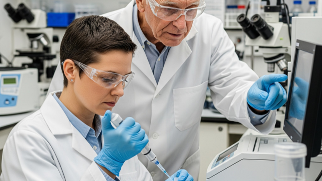
Tyrosine Hydroxylase (TH) is an important enzyme involved in the production of dopamine (a neurotransmitter that plays a key role in brain function). It helps researchers identify TH and understand diseases like Parkinson’s and other neurological disorders. It is also used in biochemistry and molecular biology.
Read this post to understand how different lab techniques are used to detect TH, ensuring accurate results in studies and experiments.
What Is Tyrosine Hydroxylase?
Tyrosine Hydroxylase (TH) is an essential enzyme that plays a key role in regulating mood and motor functions. Tyrosine hydroxylase antibody is a commonly used tool in research to identify and study this enzyme’s expression in different biological samples. TH is also a useful biomarker in the diagnosis of conditions linked to dopamine imbalances, including neurodegenerative and psychiatric disorders.
Key Aspects of Tyrosine Hydroxylase:
- Function: It controls how much dopamine, norepinephrine, and epinephrine are produced.
- Location: Found in the brain, nervous system, and adrenal glands.
- Mechanism: Uses oxygen, iron, and a cofactor called tetrahydrobiopterin to turn tyrosine into L-DOPA.
Common Lab Techniques For Tyrosine Hydroxylase Detection
Western Blotting
A lab technique which helps researchers separate proteins based on size and identify specific proteins like Tyrosine Hydroxylase (TH) using antibodies. After proteins are separated by gel electrophoresis, they are transferred to a membrane where an antibody specific to TH binds to the protein. A secondary antibody is used to identify the TH presence.
- Highly sensitive: Detects even trace amounts of TH.
- Precise: Specifically detects TH, reducing errors.
Immunohistochemistry (IHC)
This technique is used to see TH in tissue samples by using antibodies. The antibody binds to TH in tissue sections, and a color change or fluorescence signals the presence of TH.
- Shows protein location: Shows where TH is within tissues.
- Helps with mapping: Useful for studying TH spread in tissues.
Immunocytochemistry (ICC)
This technique helps researchers identify Tyrosine Hydroxylase in individual cells using antibody staining. The cells are treated with antibodies, and the location of the protein is marked.
Cell-specific analysis: Works well to study protein behavior in individual cells.
Flow Cytometry
A technique that evaluates the expression of proteins like TH in individual cells in a fluid stream. Cells are marked with fluorescent antibodies that bind to TH, and the flow cytometer measures the fluorescence intensity.
Enzyme-Linked Immunosorbent Assay (ELISA)
Researchers use this method to measure the amount of tyrosine hydroxylase in a sample using antibodies and enzyme-linked detection. A sample is added to a well coated with a TH antibody, and a color change shows the protein’s concentration.
How To Choose The Right Method For The Research?
- Sample Type:
Choose a method based on the sample (tissue, cells, or plasma) you’re studying.
For Example, IHC is great for tissue sections, whereas Western Blot works well with cell lysates or plasma.
- Resolution Requirements:
If you need to study Tyrosine Hydroxylase at a cellular or tissue level, select the right technique.
For Example, use IHC for tissue and flow cytometry or ICC for single-cell resolution.
- Quantification Needs:
Choose between qualitative or quantitative techniques.
For Example, if you’re looking for quantitative results, Western Blot or ELISA is best, whereas IHC is perfect for qualitative assessment.
Final Thought
The right identification of tyrosine hydroxylase (TH) is important for understanding dopamine-related conditions like Parkinson’s disease. By choosing the right combination of lab techniques, researchers can better set up their experiments with their research objectives.
With ongoing improvements in TH detection methods, researchers can look forward to more accurate and effective tools that will enhance the quality of their studies in the future.